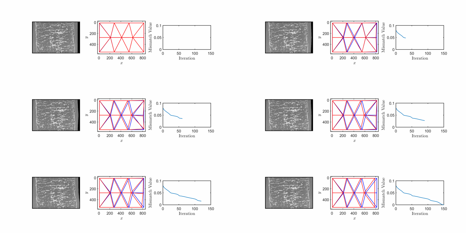
Despite comparable macroscopic compressive responses, metamaterials composed of different parent solids exhibit distinct failure mechanisms: elastomeric foams buckle via instability governed by microstructural anisotropy and polydispersity, metallic foams fold through yielding influenced by the plastic hardening of the parent solid, and carbon foams deform through progressive damage driven by stress concentrations at nodal joints.
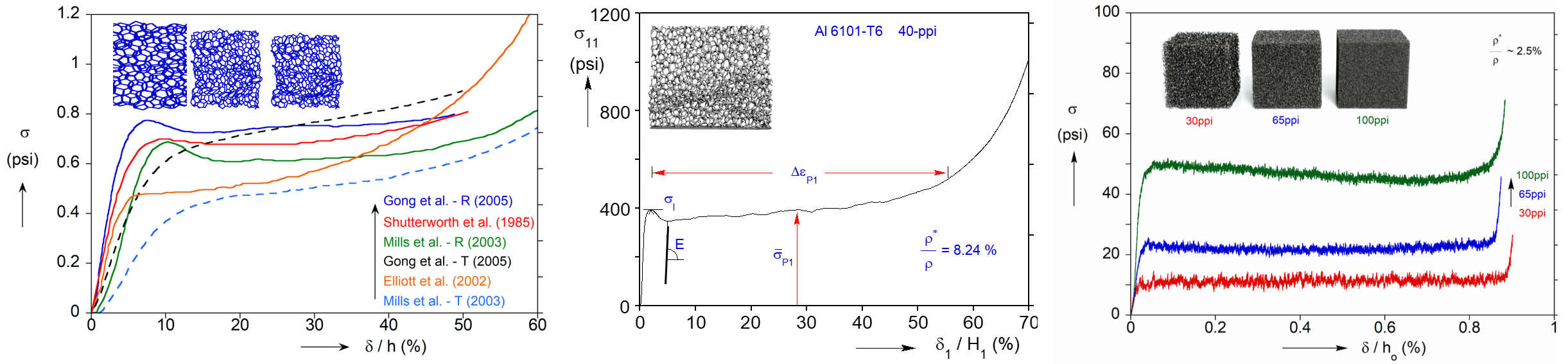
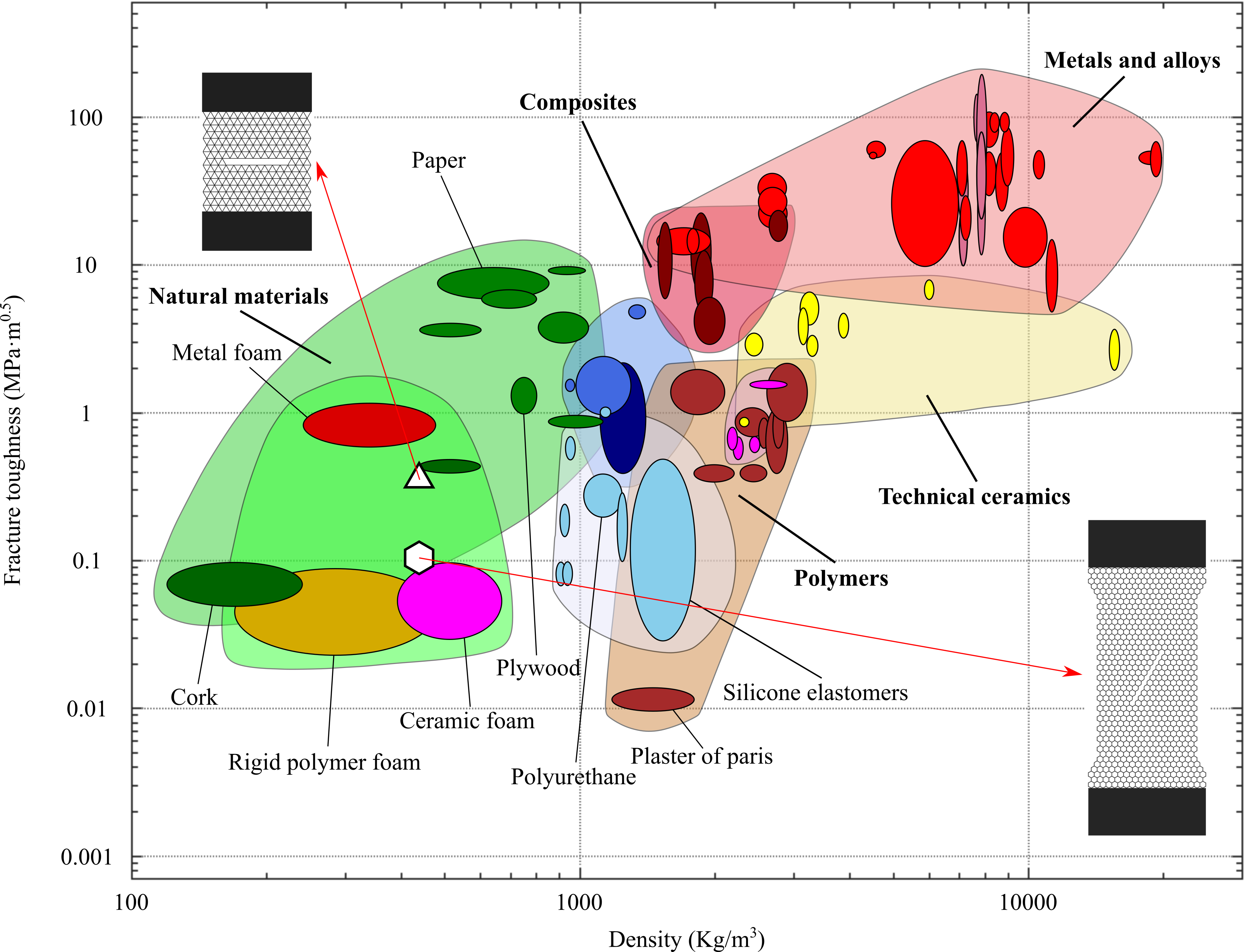
The presence of microstructure in metamaterials redistributes the stress intensity field ahead of the crack, invalidating the centric assumption of universal near crack-tip fields. Hence, the fracture mechanics of metamaterials is investigated through a global energy-based framework, and fracture energy is shown to be a more appropriate metric of fracture resistance than the conventional fracture toughness.
The dynamic performance of metamaterials is counterintuitively only minimally affected by their microstructural details and is instead governed by total mass, quasi-static strength, and the rate-dependent hardening of the parent solid. As a result, two topologies with distinct morphological characteristics but identical density and quasi-static strength exhibit similar dynamic stress levels under shock impact.
The vast number of microstructural features and their intricate interactions significantly hindered property estimation in metamaterials. Here, we generate a virtual dataset of over 8,000 ordered and disordered microstructures and develop a machine learning model for the accurate prediction of their macroscopic mechanical properties directly from their microstructures.
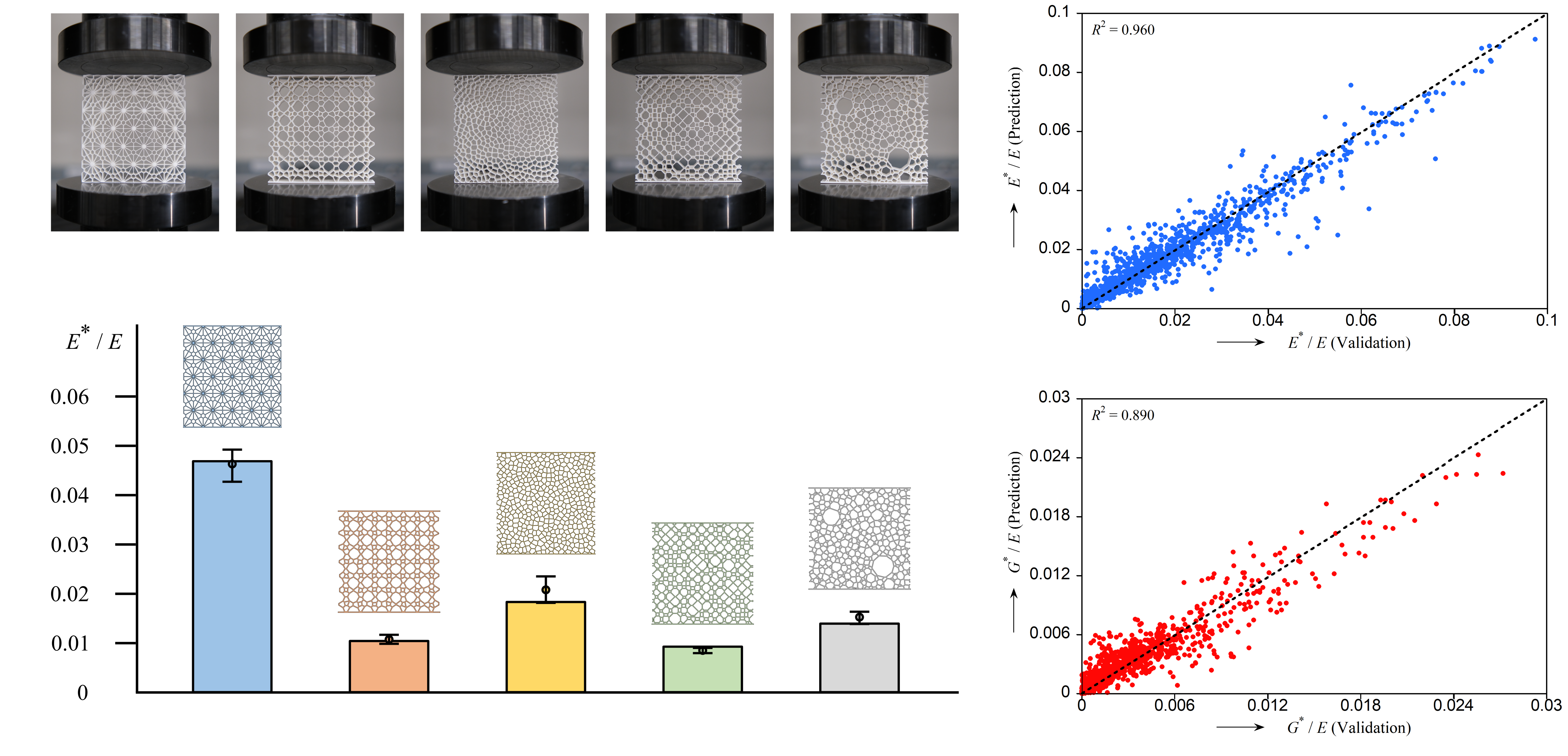
Using interpretable machine learning algorithms, we identify a clear hierarchy of microstructural features governing mechanical properties: relative density primarily modulates the overall stiffness level; nodal connectivity or cell compactness serves as secondary factor, with extreme values pushing stiffness towards the upper or lower bounds; within the intermediate regime, strut orientation becomes the decisive factor determining the final stiffness value.
• Vertebral density-modulus relationship
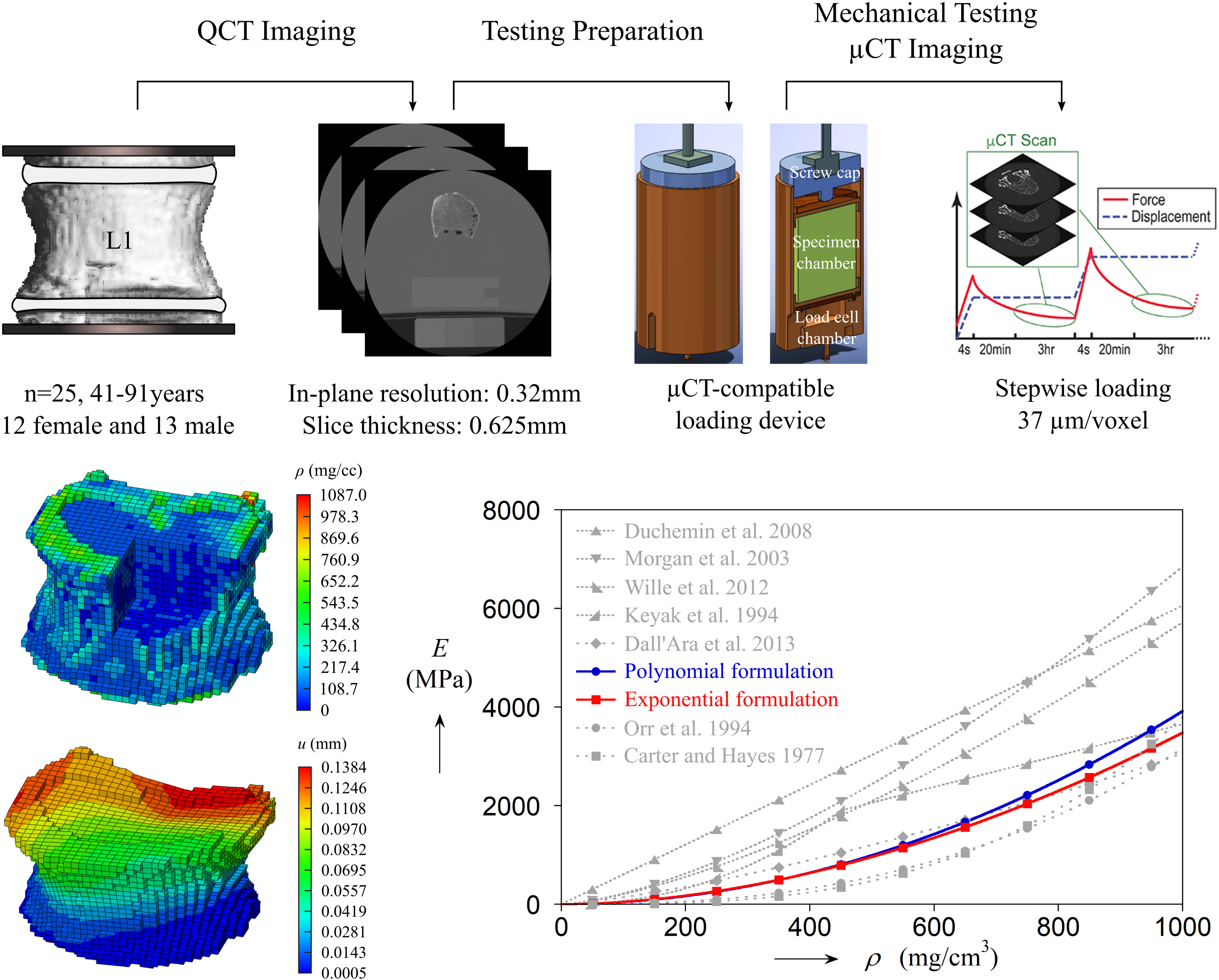
Computed tomography-based computational modeling offers a promising approach to representing bone tissue distribution; however, local structure-property mapping has largely relied on experiments that typically focus only on the centric region of the vertebra, often producing results that differ significantly. Here, we develop a data-driven framework that integrates imaging, experimental testing, and numerical modeling of human lumbar vertebrae to establish a unified density-modulus relationship across the entire vertebral body.
• Vertebral data-driven multiscale modeling
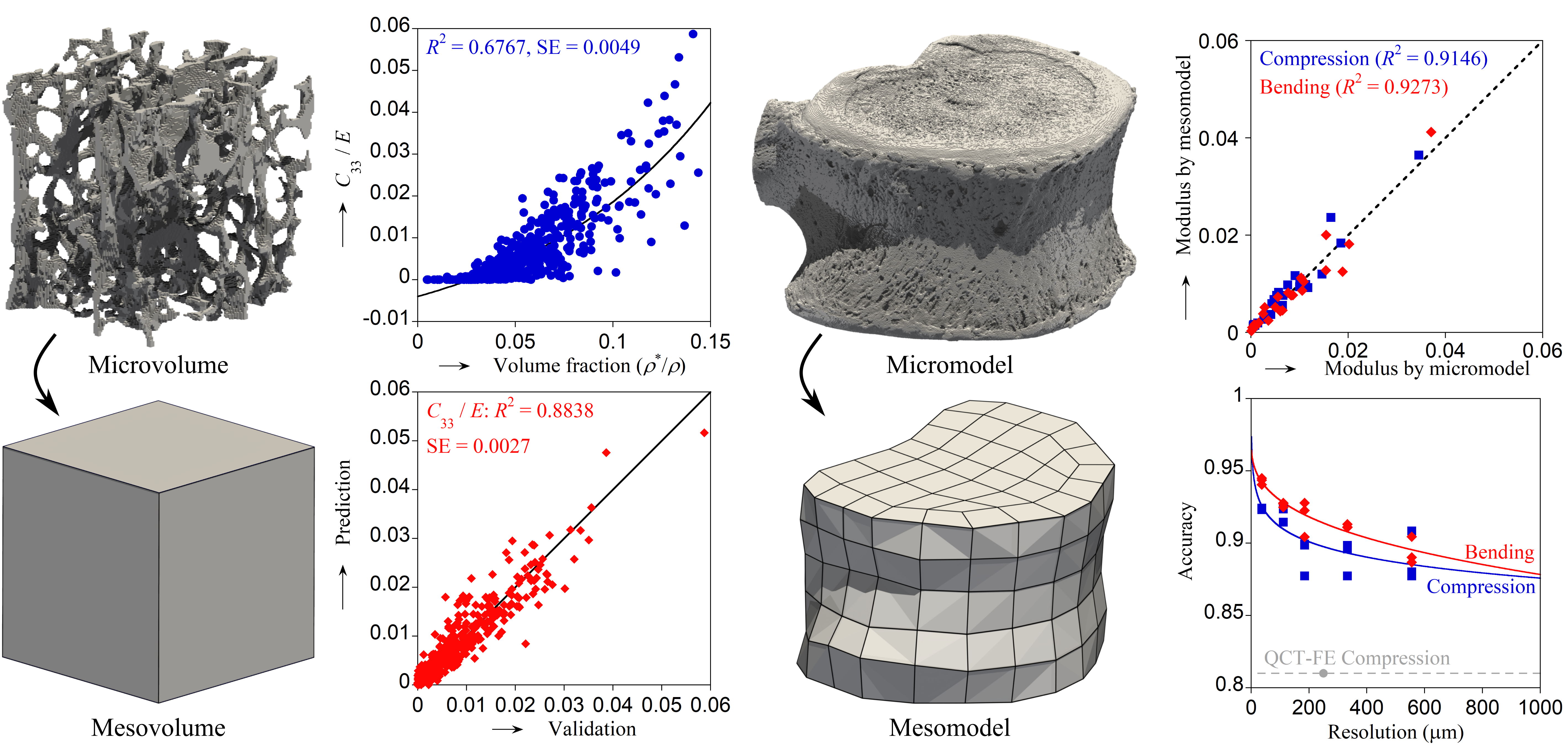
In bone biomechanics modeling, in silico models constructed from standard-resolution imaging lack adequate microstructural details and sufficient validation, whereas models built from high-resolution imaging demand excessive computational resources and prohibitively high radiation doses. Hence, we develop a data-driven multiscale modeling framework aimed at estimating human vertebral performance both accurately and efficiently, with strong potential for clinical translation.
• Software — Your DVC
To quantify deformation from continuous experimental biomedical imaging of specimens, we developed a digital volume correlation software, Your DVC. This open-source software can measure the deformation of a series of biomedical images (more than two) of a specimen, with control over interpolation order, regularization type and coefficient, and the initial field guess, enabling accurate voxel-level displacement and strain analysis.
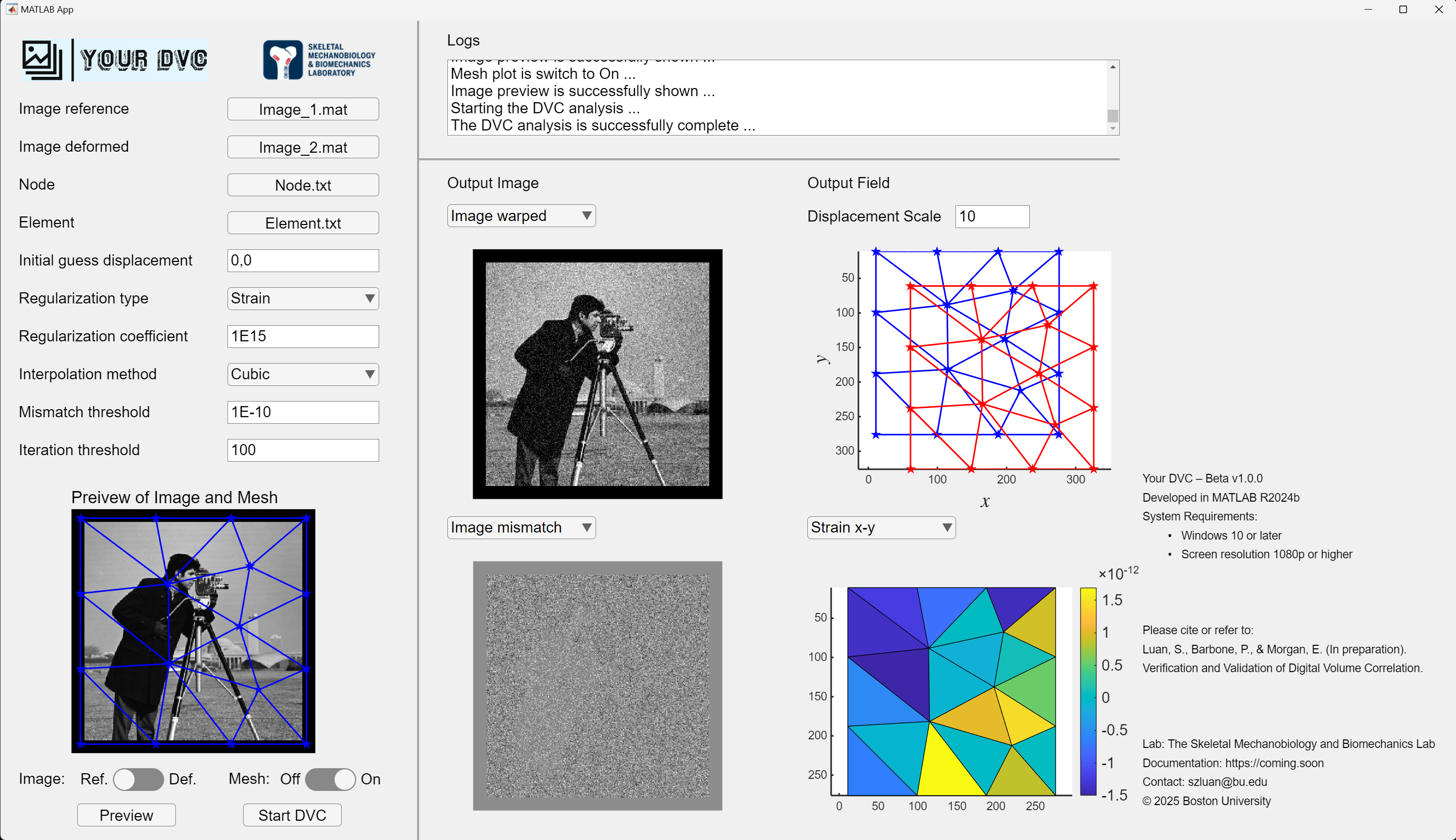
Using our software on continuous imaging data, we successfully track the experimental deformation of specimens, ranging from cellular to tissue level depending on the imaging resolution. The measured deformation can be analyzed directly or incorporated into corresponding computational models as boundary conditions or field inputs, bridging experimental observations with predictive simulations.